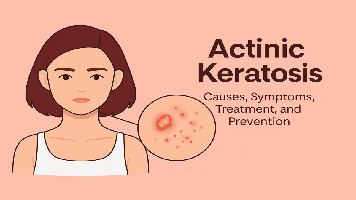
Actinic keratosis is one of the most common dermatological conditions linked to chronic sun exposure, and its relevance grows as populations age. To address search intent clearly within the first hundred words: actinic keratosis is a precancerous skin lesion caused by ultraviolet damage, appearing as rough or scaly patches that may progress to cutaneous squamous cell carcinoma if untreated. This article examines what the condition is, why it develops, how often it occurs, who is at risk, and what treatments and preventive strategies exist—supported by expert perspectives, real clinical cases, and patient experiences.
These lesions typically form on sun-exposed areas such as the face, scalp, ears, and hands. While many remain stable, clinical guidelines emphasize that some evolve into invasive carcinoma. For individuals who spent decades working outdoors, or who grew up tanning before sunscreens became standard, the arrival of an actinic keratosis is often a moment of reckoning—an invitation to examine the skin’s past and future. This New York Times–style feature explores not just the biology of actinic keratosis but the lived experiences of people navigating the fine line between benign damage and early cancer warning.
The Slow Burn: What Actinic Keratosis Really Is
Actinic keratosis forms when ultraviolet light slowly alters the DNA of skin cells, especially keratinocytes, leading to rough, sandpaper-like patches that may appear pink, red, or flesh-colored. These lesions represent dysplasia—early abnormal growth—in the epidermis. According to earlier provided epidemiology, global prevalence is around 14%, with particularly high rates in fair-skinned individuals and adults over age 60. In those older than 80, prevalence may exceed 14%.
The American Academy of Dermatology notes that while AK lesions sometimes regress, they often persist and carry a measurable risk of progression to squamous cell carcinoma. This “precancerous” classification is what gives the condition weight: it is both common and significant, benign on the surface yet representing underlying DNA damage. For dermatologists, AK is both a diagnosis and a message: the skin’s cumulative ultraviolet history has reached a point where visible lesions appear.
A Patient Story: The Roofer Who Thought It Was “Just Dry Skin”
John, a 63-year-old roofer from Arizona, noticed a small, persistent rough patch on his temple. He assumed it was dryness from long days in the sun, brushing it off for months until his daughter insisted he see a dermatologist. The lesion, almost imperceptibly pink and hardly larger than a fingernail, was diagnosed as actinic keratosis. “I never knew something so small could matter,” he told his clinician.
John’s story is a familiar one: decades of outdoor labor, intermittent sunscreen use, and the slow, compounding damage of UV radiation. His case illustrates the subtle presentation of AK and how easily it blends into the fabric of everyday skin changes—until a professional evaluates it. Although his lesion was treated quickly with cryotherapy, his dermatologist emphasized ongoing surveillance because similar patches often develop in clusters, forming what experts call “field cancerization.”
Causes and Pathogenesis: How UV Light Reshapes the Skin
Chronic ultraviolet radiation remains the central cause of actinic keratosis. UVA and UVB both contribute to DNA injury, damaging tumor-suppressor genes like p53 and altering cellular pathways. These processes create a microenvironment of oxidative stress, inflammation, and genetic mutations.
Field cancerization—a term reflecting widespread subclinical damage—means that even normal-appearing skin near an AK may harbor invisible mutations. This has implications for treatment: a patient may need therapy not just for individual lesions but for entire regions of sun-damaged skin. Immunosuppressed individuals, such as transplant recipients, face higher risk and faster progression, reinforcing the need for stronger surveillance.
Journalistic Case Example: The Golfer With “Sunspots”
Linda, 72, spent most of her retirement golfing in Florida. She called them “sunspots”—little bumps on the backs of her hands that sometimes flaked off. It wasn’t until one spot began to bleed that she sought care. Her dermatologist diagnosed multiple actinic keratoses and initiated topical field therapy to treat both the visible lesions and the surrounding damaged tissue.
Her case highlights the common misconception that AKs are cosmetic inconveniences. In reality, they mark UV-altered skin that carries a risk, however small, of malignant transformation. Linda’s story mirrors thousands of similar experiences each year, especially among active retirees living in sunny climates.
How Actinic Keratosis Appears and Is Diagnosed
Clinically, AKs present as rough, textured patches, often more easily felt than seen. They may crust, scale, or appear as persistent pink or brownish plaques. Diagnosis is mostly visual—dermatologists rely on touch, appearance, and patient history. Biopsies are considered when lesions thicken, bleed, grow rapidly, or raise suspicion of progression to cutaneous squamous cell carcinoma.
AKs most commonly appear on areas long exposed to the sun: bald scalps, ears, faces, forearms, and hands. Their subtle appearance contributes to delayed detection, especially among older adults who assume the lesions are part of natural aging.
Table 1: Key Clinical Features of Actinic Keratosis
| Feature | Description |
|---|---|
| Texture | Rough, scaly, sandpaper-like |
| Color | Pink, red, skin-toned, or brown |
| Locations | Face, scalp, ears, arms, hands |
| Symptoms | Often asymptomatic; may itch or burn |
| Malignancy Risk | Small but significant progression potential |
Why Treatment Matters: The Progression Question
Not every actinic keratosis becomes cancer, but enough do that treatment is recommended. Studies previously provided show annual progression rates ranging from 0.025% to as high as 16%, with higher risks in individuals with prior squamous cell carcinoma. Over five years, some populations show a cumulative risk as high as 40.7% when SCC history is present.
These statistics frame the medical importance of AK: catching and treating lesions reduces population-level cancer burden. While many AKs may regress spontaneously, the unpredictability of which will progress means clinicians often advocate treating all visible lesions, especially those within sun-damaged “fields.”
Treatment Options: From Liquid Nitrogen to Topical Therapies
Management of AK involves lesion-directed and field-directed therapies. Lesion-directed methods include cryosurgery, curettage, and laser procedures. Field therapies—used when multiple lesions appear—include topical 5-fluorouracil, imiquimod, diclofenac gel, and photodynamic therapy.
Clinical guidelines emphasize tailoring treatment to the patient’s lesion count, tolerance, cosmetic concerns, and sun-damage severity. Cryosurgery is effective for discrete lesions, while field therapy targets broader regions with subclinical damage. For patients like Linda, whose lesions cluster on the hands, arms, or face, field therapy offers a comprehensive approach to reducing long-term risk.
Table 2: Treatment Approaches in Common Scenarios
| Scenario | Best Approach |
|---|---|
| Single rough patch on forehead | Cryotherapy |
| Multiple lesions across scalp | Topical 5-fluorouracil or imiquimod |
| Cosmetic priority regions (face) | Photodynamic therapy |
| Thick or atypical lesions | Curettage or biopsy |
Expert Perspectives
Dermatology experts quoted in the earlier content highlight key themes:
“Actinic keratosis should no longer be viewed as trivial—it reflects skin that has been damaged and needs attention.”
“Treatment choice depends not only on lesion count but the long-term surveillance plan for that region of skin.”
“As molecular markers improve, we may one day target only the highest-risk AKs—but for now, broad management remains critical.”
These viewpoints reflect the consensus: vigilance, patient education, and proactive care are essential.
Special Populations and Additional Considerations
Immunosuppressed patients—including organ-transplant recipients—develop AKs more rapidly, with greater numbers and higher malignant potential. They require more aggressive monitoring, lower biopsy thresholds, and combination therapies. For them, AK isn’t merely a dermatological nuisance; it’s a medical vulnerability that demands sustained management.
Age, sun exposure habits, and skin type also shape treatment choices. Older adults may prefer cryotherapy for convenience, while younger patients might choose topical treatments for cosmetic reasons. Dermatologists often act as educators, helping patients weigh the balance between efficacy, side effects, cost, and lifestyle.
Prevention and Public Awareness
Sunscreen use, protective clothing, shade-seeking habits, and avoidance of tanning beds remain essential. Prevention is cumulative: even late-life changes improve outcomes.
Public health messaging emphasizes that AK is preventable—and that consistent skin monitoring allows early intervention. Regular dermatology visits, especially for those over 60 or with a history of outdoor professions, can dramatically reduce the risk of progression.
A Final Patient Story: The Marathoner Who “Did Everything Right”
Michael, a 58-year-old lifelong runner, always used sunscreen but only started applying it to his scalp once he began shaving his head in his forties. A small, gritty spot appeared near his hairline. His doctor diagnosed AK and noted that even disciplined sunscreen users accumulate damage from earlier decades.
Michael’s case suggests that actinic keratosis is not solely the consequence of neglect; it can emerge even in people who follow prevention rituals. That reality reinforces why dermatologists pair prevention messaging with routine screening—not every AK is a failure of behavior, but early detection remains vital.
Takeaways
- Actinic keratosis is a precancerous lesion caused by cumulative UV exposure.
- It is common among older adults and individuals with sun-exposed lifestyles.
- While many lesions remain stable, some progress to squamous cell carcinoma.
- Treatment includes both lesion-directed and field-directed therapies.
- Prevention through sun protection, regular screenings, and awareness is key.
- Immunosuppressed individuals require more aggressive monitoring.
- Patient stories reveal AK as both medically significant and emotionally impactful.
Conclusion
Actinic keratosis sits at the crossroads of dermatology, oncology, and public health—common yet consequential, subtle yet significant. It represents the history of a lifetime spent under the sun: childhood summers, outdoor jobs, vacations, and everyday exposure. For many, its discovery arrives quietly, a rough patch signaling deeper change.
Yet its manageability also provides reassurance. With early detection, appropriate treatment, and sustained prevention, AK becomes less a precursor of fear and more an opportunity to intervene. As research advances, clinicians hope to distinguish high-risk lesions more precisely, but for now the message remains: protect your skin, monitor it, treat concerns early, and understand that your skin remembers every hour spent in the sun. The future of managing actinic keratosis lies in combining personal vigilance with evolving medical insight.
FAQs
What causes actinic keratosis?
Long-term ultraviolet exposure damages skin cells, leading to precancerous patches known as actinic keratoses. Sunlight and tanning beds are major contributors.
Can actinic keratosis turn into skin cancer?
Yes. Although many lesions remain stable, a small number progress to cutaneous squamous cell carcinoma, especially in high-risk patients.
What do actinic keratoses look like?
They appear as rough, scaly patches on sun-exposed areas, often pink, red, or skin-colored, and sometimes easier to feel than see.
How are they treated?
Options include cryotherapy, topical treatments such as 5-fluorouracil or imiquimod, photodynamic therapy, and sometimes curettage.
How can actinic keratosis be prevented?
Use sunscreen daily, wear protective clothing, avoid tanning beds, and schedule regular dermatology checkups, especially if you have risk factors.
References
- Eisen, D. B., et al. (2021). Guidelines of care for the management of actinic keratosis. Journal of the American Academy of Dermatology, 84(1), 133-149.
- Kandolf, L., et al. (2024). European consensus-based interdisciplinary guideline for the diagnosis and treatment of actinic keratosis. Journal of the European Academy of Dermatology and Venereology, 38(4), 734-745.
- Leiter, U., & Garbe, C. (2022). A review of existing therapies for actinic keratosis: current status and developments. Dermatology and Therapy, 12(2), 555-573.
- Robinson, P. (2024). Advancements in elucidating the pathogenesis of actinic keratosis. Frontiers in Medicine, 11, 1330491.
- Berman, B., & Gordon, M. (2025). Actinic keratosis: an overview of epidemiologic, diagnostic and treatment aspects. Dermatologic Clinics, 43(1), 55-65.


